Neuronanotechnology to Cure Criminality and Mental Illness
Nancy Woolf, Ph.D.
Page 2 of 4
Now, I don’t want to argue this point too much, because there are studies that do seem to suggest long-term storage in synapses. On the other hand, it is unequivocal that dendrites continue to grow as we mature and presumably as we learn more and more.
Why should we expect information storage to occur in microtubules? For one, microtubules occupy the vast majority of space the inside of the dendrite, along with a few mitochondria [1] there aren’t too many competing sub-cellular organelles. The microtubule tracks fill up the neurons and in particular the dendrite shaft. The neurofilaments lie alongside the microtubules in the axons, but in the dendrites there is an abundance of microtubules and their associated proteins.
Information storage in microtubules also enables them to govern transport functions. There’s transport of proteins important for maintaining synaptic strength. There’s also transport of messenger RNA. Messenger RNA [2] transported in dendrites enables proteins to be translated right on the spot. I won’t go into any more detail on this.
What I am going to talk about more is the fact that there are different tubulin isotypes and that these indeed are important because they determine the binding patterns of microtubule associated proteins (MAPs). The MAPs decorate the microtubules and varying concentrations of different tubulin isotypes will produce different patterns.
What does this mean? How could the patterns along microtubules made by the MAPs translate into a mental state?
Well, before I address that directly, I want to go over the empirical data that we collected. What happens inside of neurons when animals learn? What I mean by learn is that animals show improvement on such tasks as fear conditioning, avoidance conditioning, and spatial navigation. These are well known training paradigms that are routinely used in the laboratory. We and others found that MAP2 [3], which is a microtubule associated protein, and tubulin are reorganized with memory tasks.
We showed, for example, that both the MAP2 and the tubulin proteins are proteolyzed [4]. In other words, the protein is broken down with fear conditioning. This indicated to us that what’s probably happening is the protein is broken down, then a new structure is formed, and that is the new architecture of the neuron. That would be important for memory storage.
Different laboratories replicated and extended our initial findings on MAP2 and memory. One laboratory found that the involvement of MAP2 was essential to fear conditioning. Another laboratory found that passive avoidance, a different kind of learning, reorganized the patterns of MAP2.
MAP2 is a cross-bridge. If microtubules are the sides of the ladder, then the MAPs are the rungs of a ladder. The MAPs act to strengthen the structural integrity of the microtubule matrices, but they may also play a role in transmitting and amplifying information as discussed earlier. Just as these MAP cross-bridges reorganize with learning, kinesin [5], which is a microtubule motor protein, plays a role in learning. Kinesin is a motor protein that walks along the microtubules.
Here is a picture of our data. It’s immunohistochemical data showing that in a naïve control rat, there’s little in the way of breakdown of the MAP2, whereas in these two trained rats, there is increased breakdown of the MAP2 showing up as a darker stain because broken down protein has more antigenic binding sites. We also confirmed these results with the immunoblots, which measure actual protein levels so we know that the intact protein was broken down.
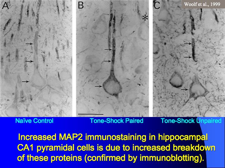
Image # 5 - MAP2
Here is an example of reorganization. These MAP2-enriched cells in a module of cerebral cortex are surrounded by regions of the cerebral cortex that show lesser amounts of this protein and lesser amounts of break down of this protein. We have observed that the modules showing enhanced staining differ from animal to animal and staining appears to be based on the most recent experience of an animal.
Let me spend a bit more time on this particular diagram. This diagram is a schematic showing the synapses involved with learning. We have the glutamate terminal, that’s the terminal that releases the neurotransmitter glutamate. And we have this
[6] acetycholine terminal, that’s the terminal that releases acetycholine.
It’s known that during learning, there is often co-release of both glutamate and acetycholine. Often there are other neurotransmitters involved, as well, but to keep this reasonable simple, I’ve limited discussion to these two.
In the spine, there are actin-filled microfilaments. These can communicate with the microtubules inside the dendrite shaft and, as I mentioned, these microtubule associated proteins, like MAP2, form bridges, but they also do more that I want to talk about.
The MAP-2 bridges attach to the microtubule and when they are phosphorylated [7], they extend outward. When the MAPs are de-phosphorylated they fold inward. Microtubules have about 43 phosphorylation sites determining how much they extend out or fold in.
The extent to which MAPs extend out or fold in provides a physical basis for a contour along the microtubule which could indeed represent information. This essentially is the idea that I’m developing, that we could have a contour that would represent information inside of the cell that would then coordinate with housekeeping functions like transporting receptors to synapses. This physical contour would be capable of transmitting and amplifying information in the form of electromagnetic waves.
How could these proteins be phosphorylated with learning or synaptic activation? During learning we have activation at both glutamate and acetylcholine terminals leading to more neurotransmitter release. After these neurotransmitters bind with their receptors, they activate second messengers, which in turn phosphorylate microtubule proteins, such as MAP2.
And then there is conductive signaling along microtubules, which will be affected by the phosphorylation of MAPs. Rather than microtubules being mere structural entities or even transports tracks, they may transmit information according to their semi-conductive properties.
I also mentioned there are different tubulin isotypes, especially in the brain. But why are there so many isotypes in the brain? Most cells in the body only have the most prevalent tubulin isotype. Beta-1, for example, is a tubulin isotype that’s found in all cells. There are rarer Beta-2, Beta-3, Beta-4, and [Beta-6] tubulin isotypes that are specifically found in brain. Why?
Footnotes
[1] Mitochondria - Mitochondria provide the energy a cell needs to move, divide, produce secretory products, contract - in short, they are the power centers of the cell.http://www.cellsalive.com/cells/mitochon.htm
March 6, 2007 2:58 PM EST
[2] RNA – Ribonucleic acid; a polymeric constituent of all living cells and many viruses, consisting of a long, usually single-stranded chain of alternating phosphate and ribose units with the bases adenine, guanine, cytosine, and uracil bonded to the ribose. The structure and base sequence of RNA are determinants of protein synthesis and the transmission of genetic information.
Stedman, The American Heritage Medical dic·tion·ar·y, Boston, New York: Houghton Mifflin Company, 2004: 719.
[3] MAP2 - This gene encodes a protein that belongs to the microtubule-associated protein family. The proteins of this family are thought to be involved in microtubule assembly, which is an essential step in neurogenesis. The products of similar genes in rat and mouse are neuron-specific cytoskeletal proteins that are enriched in dentrites, implicating a role in determining and stabilizing dentritic shape during neuron development. A number of alternatively spliced variants encoding distinct isforms have been described.
http://www.ncbi.nlm.nih.gov/entrez/query.fcgi
&
http://www.gene.ucl.ac.uk/nomenclature
March 6, 2007 3:07 PM EST
[4] Proteolysis – The hydrolytic break-down of proteins into simpler, soluble substances, as occurs in digestion.
Stedman, The American Heritage Medical dic·tion·ar·y, Boston, New York: Houghton Mifflin Company, 2004: 673.
[5] Kinesin - the founding member of a superfamily of microtubule-based ATPase motors that perform force-generating tasks such as organelle transport and chromosome segregation.
[6] Acetycholine - often abbreviated as ACh, was the first neurotransmitter to be identified. It is a chemical transmitter in both the peripheral nervous system (PNS) and central nervous system (CNS) in many organisms including humans. Acetylcholine is the neurotransmitter in all autonomic ganglia.http://en.wikipedia.org/wiki/Acetylcholine
March 6, 2006 4:12 PM EST
[7] Phosphorylation – the addition of phosphate to an organic compound through the action of a phosphorylase or kinase.Stedman, The American Heritage Medical dic·tion·ar·y, Boston, New York: Houghton Mifflin Company, 2004: 629.
1 2 3 4 Next Page>